Author: Julie Graham

Oral Surgery Dental Code

Oral surgery is a specialized branch of dentistry that focuses on surgical procedures within the mouth, jaw, and surrounding areas. To streamline billing and provide accurate treatment information, dental professionals rely on specific coding systems, such as the oral surgery dental code. This coding system allows oral surgeons to document procedures, treatments, and consultations accurately. In this article, we’ll explore everything you need to know about the oral surgery dental code, from understanding its structure to using it effectively in your practice. Whether you’re a dentist, oral surgeon, or insurance professional, this guide is packed with insights to make coding easier and more efficient.

What is Oral Surgery Dental Code?

Oral surgery dental codes are a set of standardized codes used by dentists and oral surgeons to describe surgical procedures performed on the oral cavity, jaw, and facial structures. These codes, commonly referred to as CPT codes (Current Procedural Terminology), are essential for documentation, insurance claims, and billing.

Why Oral Surgery Codes Are Important:

Accurate coding ensures proper reimbursement from insurance providers and helps avoid misunderstandings during claims. These codes are updated periodically to reflect new surgical techniques or changes in medical practice.

Where Are These Codes Used?

Oral surgery codes are primarily used in medical records, insurance billing systems, and for healthcare professionals to communicate surgical procedures across platforms.

Key Features of Oral Surgery Codes:

They usually consist of a series of digits, each representing a specific procedure or treatment within the field of oral surgery.

Importance of Correctly Using Oral Surgery Dental Codes

Properly using oral surgery dental codes is crucial for ensuring smooth billing and reimbursement processes. If a dental code is used incorrectly, it can lead to claim rejections, delayed payments, or even legal issues. Below are the key reasons why using the right code is important:

Accurate Reimbursement:

Correct codes allow dental professionals to receive the appropriate compensation for services rendered.

Compliance with Insurance Requirements:

Different insurance providers may require specific codes for processing claims. Understanding which code to use can prevent errors in the claim process.

Efficient Documentation:

Codes help streamline patient records, making it easier for healthcare providers to track treatments and procedures over time.

Types of Oral Surgery Codes

There are several types of oral surgery codes, each designed for specific surgical procedures or services. These codes are generally categorized as follows:

Simple Extractions:

These codes are used for basic tooth extractions that do not require complex surgical intervention.

Complex Extractions:

These codes cover more intricate tooth extractions, such as the removal of impacted teeth.

Jaw Surgery (Orthognathic Surgery):

Used for surgical procedures aimed at correcting jaw deformities or misalignments.

Implant Surgery:

Implant-related codes are used when a dental implant is placed into the jawbone.

Biopsy Codes:

These codes are used for procedures that involve taking a sample of tissue for examination.

Commonly Used Oral Surgery Codes

Below is a list of some of the most commonly used oral surgery dental codes:

  • CPT Code 41899: Unspecified oral surgery

  • CPT Code 77402: Maxillary sinus surgery

  • CPT Code 78106: Maxillofacial prosthesis placement

  • CPT Code 70110: Biopsy of oral tissue

These are just a few examples. To get a full list of oral surgery codes, it’s essential to refer to official coding guides and resources.

How to Use Oral Surgery Dental Codes Effectively

Using oral surgery dental codes effectively requires understanding the nuances of the coding system and staying up to date with any changes or updates. Below are some tips for efficient usage:

Stay Updated with Code Changes:

Dental codes are updated regularly, so it’s essential to stay informed about the latest versions and modifications.

Use Code Lookup Tools:

Various online platforms and software tools can help you search for and verify codes.

Consult the Official Coding Manuals:

Ensure you’re using the right code by consulting official resources like the Current Procedural Terminology manual or the American Dental Association’s (ADA) Code on Dental Procedures and Nomenclature.

Ensure Accuracy in Documentation:

Every surgery and procedure should be well-documented. The correct code reflects the exact nature of the procedure performed.

Avoid Using General Codes:

Always opt for the most specific code available to ensure accurate reporting and billing.

Common Mistakes in Oral Surgery Dental Coding and How to Avoid Them

While using oral surgery dental codes, there are several common mistakes that can lead to issues with insurance claims or patient records. These include:

Using Outdated Codes:

Always verify that the code you’re using is up-to-date to avoid claim rejections or issues with insurance providers.

Misclassifying Procedures:

Sometimes, a procedure may seem simple, but it’s more complex than anticipated. Ensure that you use the correct code for complex surgeries.

Incorrect Documentation:

Inaccurate or incomplete documentation can lead to errors in code selection. Make sure every procedure is properly recorded.

Overcoding or Undercoding:

Overcoding may lead to accusations of fraud, while undercoding can result in lower reimbursement.

FAQs:

What is the Oral Surgery Dental Code used for?
Oral surgery dental codes are used to document and bill for surgical procedures performed on the oral cavity and surrounding areas.

How often are oral surgery codes updated?
Oral surgery codes are typically updated annually, but they can change more frequently depending on advancements in medical practice.

What happens if I use the wrong oral surgery dental code?
Using the wrong code can lead to claim rejections, delayed payments, and potential legal issues.

Where can I find the full list of oral surgery codes?
The full list of oral surgery codes can be found in the official CPT coding manual and ADA’s Code on Dental Procedures and Nomenclature.

Can I use the same code for different types of oral surgeries?
No, each type of oral surgery has its own unique code. Using the correct code ensures accurate billing and patient documentation.

Conclusion

Mastering the oral surgery dental code system is essential for any dental or medical practice. Accurate coding ensures proper reimbursement, compliance with insurance requirements, and the maintenance of detailed patient records. By understanding the different types of codes, using them correctly, and avoiding common mistakes, dental professionals can enhance their practice’s efficiency and reduce the risk of errors in billing.

How to Make a Dental Bridge at Home

A dental bridge is a great solution for missing teeth, offering both functional and aesthetic benefits. While dental professionals typically handle the creation and placement of bridges, many people wonder if it’s possible to create one at home. This article will walk you through the entire process of making a dental bridge at home, using accessible materials and simple techniques. If you’re considering this as a temporary solution, it’s essential to understand the process and the precautions you should take to ensure both safety and effectiveness. Let’s dive into the steps, materials, and tips for creating a dental bridge at home.

What You Need to Know About Dental Bridges

Before diving into the DIY process, let’s take a moment to understand what a dental bridge is and how it functions. A dental bridge is a prosthetic device designed to fill the gap left by one or more missing teeth. It typically involves anchoring the bridge to adjacent teeth or implants. Making one at home is not an ideal solution for long-term use, but it can serve as a temporary fix if you’re in between dental appointments.

Materials You’ll Need to Make a Dental Bridge at Home

Creating a dental bridge at home requires certain materials. Here’s a list of what you’ll need:

  • Dental impression material (for making molds)

  • Temporary bridge material (such as dental wax or resin)

  • Scissors or cutters

  • A mold tray

  • A mirror for placement and fitting

Tip: Always ensure that the materials you use are safe for oral use, and never attempt to make a dental bridge from materials that could harm your gums or teeth.

Step-by-Step Instructions for Making a Dental Bridge at Home

1. Create a Mold of Your Teeth

Start by creating an impression of the teeth on either side of the gap. Use dental impression material to make the mold, following the manufacturer’s instructions carefully. This will help ensure the bridge fits securely once completed.

2. Prepare the Temporary Bridge Material

Once the mold is ready, prepare the temporary bridge material. Dental wax or resin can be shaped into the desired form and used to fill in the gap. Be sure to shape the material to mimic the appearance of your natural teeth.

3. Fit the Temporary Bridge

Place the temporary bridge carefully into the gap, checking that it fits well with your natural teeth. You might need to adjust the size slightly to ensure comfort and proper alignment.

4. Secure the Bridge

To keep the bridge in place, use a temporary dental adhesive. Apply a small amount of adhesive to the underside of the bridge, then press it into position. Make sure the bridge is not too tight or loose, as this could cause discomfort or affect the fit.

5. Allow the Bridge to Set

Let the temporary bridge set in place for the recommended amount of time. Once it’s secure, you should be able to use the bridge as a functional solution until you can visit a dental professional.

Common Mistakes to Avoid When Making a Dental Bridge at Home

  • Using inappropriate materials: Always use dental-grade materials to avoid irritation or damage.

  • Improper fit: If the bridge is too tight or loose, it could cause discomfort or lead to gum irritation.

  • Failure to secure the bridge properly: If the bridge isn’t securely fixed, it can fall out, leading to potential choking hazards.

How to Maintain Your DIY Dental Bridge

To keep your temporary dental bridge in good condition, follow these care tips:

  • Avoid eating hard or sticky foods that might dislodge the bridge.

  • Clean the bridge and surrounding area gently with a soft toothbrush and toothpaste.

  • Be mindful of any discomfort or changes in the fit, and seek professional help if needed.

Frequently Asked Questions (FAQs)

1. Can I make a permanent dental bridge at home?

No, making a permanent dental bridge at home is not recommended. Permanent bridges require professional care and materials to ensure they fit properly and are safe for long-term use.

2. How long can I keep a homemade dental bridge in place?

A homemade dental bridge should only be used temporarily. It can last anywhere from a few days to a couple of weeks, but it’s important to visit a dentist for a permanent solution as soon as possible.

3. Is it safe to make a dental bridge at home?

While it’s generally safe to make a temporary dental bridge at home, it is important to use appropriate materials and ensure the fit is correct. Consult with a dentist if you experience any discomfort or issues.

4. What materials are used for a homemade dental bridge?

Common materials include dental wax, resin, and impression material. Always use materials that are specifically designed for dental use to avoid potential harm.

5. Can I eat normally with a DIY dental bridge?

It’s best to avoid hard, sticky, or chewy foods while wearing a DIY dental bridge. Stick to softer foods and be mindful of the fit to avoid dislodging it.

Conclusion

While making a dental bridge at home can be a helpful temporary fix, it’s important to remember that this is not a long-term solution. Always consult with a dentist to ensure you receive professional care. This DIY method should only be used as a stopgap measure until you can receive proper dental treatment. Making a dental bridge at home can save you from discomfort in the short term, but professional care is the best option for maintaining long-term oral health.

Dental Code for Bone Graft: Proper Billing and Reimbursement

Bone grafts are essential in dental procedures, particularly when a patient lacks sufficient bone for implants or other restorative work. The dental code for bone graft plays a crucial role in ensuring that these procedures are properly coded for insurance and billing purposes. Understanding these codes is essential for dental professionals and patients alike to navigate the complexities of insurance claims and reimbursement. In this article, we’ll dive deep into the details of dental codes for bone grafting, why they matter, and how they impact dental treatment plans.

What Is the Dental Code for Bone Graft?

Bone grafting is commonly used in dental implant surgeries when a patient has insufficient bone structure to support the implant. The dental codes for these procedures help standardize treatment, making it easier for dental professionals to bill and insurance companies to process claims.

The dental code for bone grafting varies based on the type of graft performed. There are codes for autografts, allografts, and xenografts, with specific numbers assigned to each one. Understanding these codes is crucial for both accurate billing and ensuring that the patient receives the proper reimbursement.

Key Codes to Know:

  • D7953 – Bone Replacement Graft

  • D7950 – Bone Graft Procedure

  • D4263 – Bone Grafting in Conjunction with Guided Tissue Regeneration

It’s important for dental practices to stay updated on these codes to avoid errors and ensure proper reimbursement.

Key Benefits of Bone Graft Dental Codes:

  • Insurance Reimbursement: Accurate dental codes ensure that patients are reimbursed for bone graft procedures.

  • Standardization: These codes provide a universal language for dental professionals, ensuring consistency across the industry.

  • Efficient Billing: Correct coding reduces delays in payment and minimizes the risk of claims being denied.

  • Improved Patient Experience: Clear understanding of the process reduces confusion for patients regarding insurance and payment.

Bone grafting is often necessary for dental implants, making these codes vital for both patient care and practice management.

The Different Types of Bone Grafts and Their Codes

Understanding the different types of bone grafts is essential for accurate coding. Here are the most common types of bone grafts and their corresponding codes:

  1. Autograft (D7953):

    • A graft taken from the patient’s own body, often from the chin or the back of the jaw.

  2. Allograft (D7950):

    • A bone graft harvested from a donor, typically a cadaver, and treated for safety.

  3. Xenograft (D4263):

    • A graft from a non-human species, commonly bovine (cow) bone, used when other graft materials aren’t available.

Each type of bone graft serves a unique purpose and is coded differently for insurance purposes.

How to Use the Dental Code for Bone Graft in Your Practice

Best Practices for Accurate Coding:

  • Stay Informed: Keep up to date with the latest code changes and updates from the American Dental Association (ADA).

  • Training: Regular training for staff members on dental codes and billing practices.

  • Consultation: Consult with an experienced dental coder if unsure about the correct code.

  • Documentation: Ensure proper documentation of the bone graft procedure to support your claims.

Accurate coding will prevent claim denials and ensure your practice gets paid for the work performed.

Potential Challenges and Common Mistakes with Bone Graft Coding

Challenges to Watch Out For:

  • Incorrect Code Selection: Selecting the wrong dental code can lead to claim rejections or delays.

  • Outdated Codes: Using outdated codes for newer procedures can cause issues with reimbursement.

  • Lack of Documentation: Failing to provide the correct documentation can result in denied claims.

To minimize these challenges, it’s essential for dental offices to have a system in place to double-check codes and ensure accuracy.

Frequently Asked Questions (FAQs):

1. What is the most commonly used dental code for bone grafting?

The most common dental code for bone grafting is D7953, which refers to bone replacement graft procedures. However, the specific code may vary depending on the type of graft used.

2. How does accurate coding affect insurance reimbursement?

Accurate coding ensures that insurance companies can process claims without delays. Incorrect codes can lead to denied claims or a lack of reimbursement, affecting both the dental practice and the patient.

3. Can I use the same code for all types of bone grafts?

No, different types of bone grafts require different codes. Autografts, allografts, and xenografts each have their own specific codes.

4. Are dental codes for bone grafts updated regularly?

Yes, dental codes are updated periodically by the ADA to reflect new procedures and advancements in dental technology. It’s important for dental offices to stay up-to-date with these changes.

5. What should I do if a bone graft procedure gets rejected by insurance?

If a bone graft procedure is rejected, review the documentation and ensure the correct code was used. If needed, resubmit with the necessary supporting information or consult with an insurance claims expert.

Conclusion:

In conclusion, understanding and correctly using the dental code for bone graft is vital for efficient billing and insurance claims processing. By staying informed about the latest codes, types of grafts, and best practices for coding, dental professionals can avoid delays and ensure patients receive the correct reimbursement. Accurate coding is not just important for financial aspects of dental practices but also for maintaining patient satisfaction and trust.

Does Caffeine Affect Novocaine? Its Impact on Dental Procedures

Caffeine is one of the most consumed substances worldwide. For many, a cup of coffee in the morning is an essential part of their routine. However, if you’re about to undergo a dental procedure involving novocaine, you might be wondering how your caffeine intake could impact the effectiveness of the anesthesia. In this article, we’ll explore the science behind novocaine, how caffeine affects it, and why it’s crucial to be mindful of your caffeine consumption before a dental visit.

What is Novocaine and How Does It Work?

Novocaine (procaine) is a local anesthetic commonly used in dental procedures to numb specific areas of the mouth. It blocks nerve signals, preventing pain in the treated area. Novocaine is injected directly into the gums or tissue near the targeted area, and it typically begins to take effect within minutes. It works by inhibiting sodium channels, which are essential for nerve signal transmission. This makes it a highly effective method of preventing discomfort during dental procedures like fillings, extractions, and root canals.

However, there are several factors that can impact how well novocaine works. One of the most significant of these is caffeine, which can alter the effectiveness of novocaine and make dental procedures less comfortable.

How Caffeine Affects Novocaine’s Effectiveness

Caffeine has stimulating properties that can interfere with the numbing effects of novocaine. Here’s how caffeine can affect the anesthetic:

Caffeine and Blood Flow:

Caffeine is a vasoconstrictor, which means it constricts blood vessels. When you consume caffeine, your blood vessels narrow, reducing blood flow to specific areas, including the site of the novocaine injection. As a result, the anesthesia may not be absorbed as efficiently, which can reduce its effectiveness. The slower the absorption, the less effective the numbing will be.

Faster Metabolism of Novocaine:

Another factor to consider is how caffeine can increase the metabolism of drugs in your system, including novocaine. Caffeine is known to speed up your body’s ability to break down substances, which means it can cause novocaine to be metabolized faster. This results in the anesthesia wearing off more quickly, making it necessary for your dentist to use more anesthetic to maintain the desired level of numbness throughout the procedure.

Increased Sensitivity to Pain:

Caffeine stimulates the central nervous system, which can increase sensitivity to pain. Although caffeine doesn’t directly cause pain, its stimulating effect on the nervous system can amplify your perception of discomfort, making you feel more sensitive during the dental procedure. This can make the numbing effect of novocaine seem less potent.

The Impact of Caffeine on Your Dental Appointment

For individuals who drink caffeinated beverages regularly, the effects of caffeine on novocaine can be significant. Here’s a deeper look into how caffeine consumption might influence your dental visit.

Should You Avoid Caffeine Before Your Appointment?

It’s generally recommended to avoid caffeine for at least 6 hours before your dental procedure. This ensures that the novocaine has the best chance of working effectively. If you consume caffeine too close to your appointment, you may experience reduced anesthesia, leading to discomfort during the procedure.

How Much Caffeine Is Too Much?

Even moderate caffeine consumption can potentially affect the outcome of your dental procedure. If you’re someone who drinks coffee regularly, you don’t have to completely cut it out, but it’s essential to monitor your intake. A single cup of coffee or an energy drink might not drastically affect novocaine, but anything beyond that could.

Should You Consult Your Dentist About Caffeine?

Always inform your dentist about your caffeine habits, especially if you regularly consume large amounts before a visit. Your dentist may recommend adjusting the dosage of novocaine or advise you to avoid caffeine altogether prior to the procedure.

Why Does Caffeine Have These Effects?

Understanding the science behind caffeine’s interaction with novocaine can help clarify why it’s important to avoid it before dental procedures.

Caffeine as a Vasoconstrictor:

Caffeine works by constricting blood vessels. While this is helpful in some medical contexts, it can make it harder for novocaine to be absorbed by the tissues in the area being treated. As a result, the local anesthetic may not be able to numb the area as effectively, leading to a less-than-optimal experience during your procedure.

Caffeine’s Impact on the Nervous System:

Caffeine is a stimulant that affects the central nervous system. This can interfere with the way your body responds to novocaine. While novocaine numbs the nerves in a targeted area, caffeine can make your nervous system more reactive, potentially reducing the effectiveness of the anesthetic.

Increased Metabolism of Novocaine:

Caffeine can speed up the metabolism of novocaine in your system. As your body processes the anesthetic more quickly, the numbing effect fades sooner than anticipated, which may lead to a less comfortable procedure, especially during longer treatments.

Frequently Asked Questions (FAQs)

Q1: Can I drink coffee before getting a dental injection?
A: It’s best to avoid caffeine before a dental injection as it can reduce the effectiveness of novocaine.

Q2: How long before a dental appointment should I avoid caffeine?
A: Aim to avoid caffeine for at least 6 hours before your dental procedure for optimal results.

Q3: Will one cup of coffee affect novocaine?
A: One cup of coffee is unlikely to drastically affect novocaine, but it’s still advisable to limit caffeine intake before your appointment.

Q4: Can novocaine wear off faster if I drink caffeine?
A: Yes, caffeine can cause your body to metabolize novocaine faster, leading to a quicker loss of numbness.

Q5: Should I inform my dentist if I’ve consumed caffeine?
A: Yes, always inform your dentist about your caffeine consumption so they can adjust the anesthetic dosage if necessary.

Conclusion

In conclusion, caffeine can indeed affect the effectiveness of novocaine during dental procedures. By constricting blood vessels, increasing metabolism, and stimulating the nervous system, caffeine can reduce the potency of novocaine, leading to potential discomfort during treatment. To ensure that your dental appointment goes smoothly, it’s advisable to avoid caffeine for at least 6 hours before the procedure. Always consult your dentist if you have any concerns about your caffeine consumption and its impact on your dental anesthesia.

Crown Lengthening Dental Code: Everything You Need to Know

Crown lengthening is a dental procedure that improves the health and appearance of your teeth by reshaping the gums. It’s essential for several dental treatments, and knowing the crown lengthening dental code helps both patients and dental professionals navigate insurance claims and procedural requirements. This comprehensive guide will explore everything about crown lengthening, from its benefits to the dental code you need to know. Whether you’re considering this procedure for cosmetic reasons or as part of a treatment plan, understanding the dental code is crucial for ensuring smooth medical insurance processing and a successful outcome.

Crown Lengthening Dental Code: What It Is and Why It Matters

Crown lengthening is primarily done when there is insufficient tooth structure above the gum line to restore or repair a tooth. The dental code for this procedure ensures that it is correctly documented for billing and insurance purposes.

In many cases, this procedure is required for:

  • Cosmetic enhancement: Giving a more proportionate smile.

  • Restorative work: Ensuring proper dental restoration by exposing more of the tooth.

  • Health-related concerns: Improving gum health and preventing infection.

The crown lengthening dental code allows the dental practice to classify this procedure within their system, which is essential for efficient communication with insurance companies and accurate billing.

What is the Crown Lengthening Dental Code?

The crown lengthening dental code refers to the specific numeric code used to document the procedure for billing and insurance purposes. The CPT code for crown lengthening typically falls under a series of codes related to oral surgery, especially those dealing with gingival resection and tooth exposure.

How Crown Lengthening Improves Your Smile and Health

  • Enhances Smile Aesthetics: Crown lengthening can create a more balanced smile by reducing excess gum tissue.

  • Facilitates Restorative Treatments: It allows for the proper fitting of crowns or bridges when there isn’t enough tooth structure exposed.

  • Improves Oral Health: The procedure helps prevent gum diseases that can develop in hard-to-reach areas.

These benefits demonstrate why crown lengthening is often considered essential for both cosmetic and functional purposes.

Navigating Insurance with the Crown Lengthening Dental Code

When crown lengthening is necessary, understanding the crown lengthening dental code is vital for insurance claims. Insurance companies require these codes to verify procedures and determine reimbursement. By using the right codes, dental professionals ensure that their services are covered under the patient’s insurance plan.

  • Proper Code Usage: Ensures efficient billing and reduces the chances of claim rejections.

  • Explaining the Need for the Procedure: Clear use of the crown lengthening dental code can help clarify the medical necessity of the treatment.

What Are the Costs Involved with Crown Lengthening?

  • Procedure Costs: On average, crown lengthening can cost between $200 and $3,000, depending on the complexity and the dental office’s location.

  • Insurance Coverage: With the right crown lengthening dental code, patients can be reimbursed for part or all of the cost, depending on their insurance plan.

hen Crown Lengthening is Part of a Larger Treatment Plan

Crown lengthening is often a necessary step in procedures like dental crowns, bridges, or implants. By reshaping the gums and exposing more of the tooth, it allows for better fitting restorations and helps avoid complications that may arise from insufficient tooth structure.

FAQs

What is the crown lengthening dental code?

The crown lengthening dental code is a unique code used to document the procedure for insurance and billing purposes. It helps dental professionals communicate with insurers and ensures that the treatment is covered.

How does crown lengthening improve my smile?

It removes excess gum tissue, providing a more balanced and aesthetically pleasing smile. This is often referred to as a “gummy smile” treatment.

Is crown lengthening covered by insurance?

Depending on the insurance provider, crown lengthening may be covered if it’s deemed medically necessary, such as when preparing for restorative dental work like crowns or bridges.

How long does the crown lengthening procedure take?

The procedure typically lasts between one to two hours, depending on how much tissue needs to be reshaped.

What should I expect during recovery from crown lengthening?

Recovery can take a few weeks, during which patients are advised to avoid hard foods, follow a prescribed oral care routine, and schedule follow-up appointments to monitor healing.

Conclusion

Crown lengthening is a versatile and beneficial dental procedure, whether for cosmetic, restorative, or health-related reasons. Understanding the crown lengthening dental code ensures that the procedure is accurately documented, covered by insurance, and properly billed. Whether you’re a patient or a dental professional, knowing this code helps streamline the process and ensures smoother dental care and financing.

What Does a Tooth Look Like After Root Canal

Root canal treatment is one of the most common dental procedures performed worldwide, yet many patients have little understanding of how their tooth will appear afterward. When your dentist recommends a root canal, they’re essentially saving your natural tooth from extraction while removing the infected or damaged pulp inside. This procedure has evolved significantly over the years, with modern techniques focusing not just on functionality but on maintaining the natural appearance of your tooth as well.

The external appearance of a tooth immediately after a root canal often surprises patients. In most cases, the tooth looks remarkably similar to how it did before the procedure, especially if a temporary filling is placed. However, there are subtle changes that occur both immediately and over time that patients should understand. These changes are normal and typically don’t affect the tooth’s function or your smile aesthetics, especially with proper restoration.

The tooth undergoes significant transformation during root canal therapy. The dentist removes all the living tissue (pulp) from inside the tooth, including the nerve and blood vessels. This space is then cleaned, shaped, disinfected, and filled with a rubber-like material called gutta-percha. While this internal change doesn’t directly affect the external appearance, it does make the tooth more brittle over time, which is why most dentists recommend placing a crown after the root canal procedure.

Patients often worry that their tooth will look “dead” or noticeably different after a root canal. This misconception couldn’t be further from the truth. With modern dental techniques and materials, root canal-treated teeth can blend seamlessly with your natural smile. The key factors that determine the final appearance include the tooth’s initial condition, the extent of damage before treatment, the quality of the root canal procedure, and the type of restoration placed afterward.

The journey of a tooth through root canal treatment and restoration is fascinating from both a biological and aesthetic perspective. Understanding what to expect can alleviate anxiety and help patients make informed decisions about their dental care. As we explore the various aspects of post-root canal tooth appearance, you’ll gain valuable insights into this common yet often misunderstood dental procedure.

The Root Canal Procedure: A Brief Overview

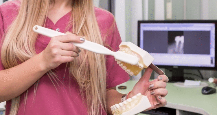
A root canal procedure, also known as endodontic therapy, is designed to save a tooth that has become infected or severely decayed. During this treatment, your dentist or endodontist will first administer local anesthesia to ensure your comfort. They then create a small opening in the crown of the tooth to access the pulp chamber. Using specialized instruments, they carefully remove the infected or damaged pulp, clean and shape the canals, and then fill them with gutta-percha. Finally, the access point is sealed with a temporary or permanent filling.

Immediate Post-Procedure Appearance

Immediately after your root canal, your tooth will look much the same as it did before the procedure, with one notable difference: the presence of a temporary filling. This filling is typically made of a soft material that’s easy for your dentist to remove when you return for your permanent restoration. The tooth itself might feel slightly different due to the absence of nerve tissue, but visually, it should blend in with your surrounding teeth. Some patients notice minimal swelling around the treated area, but this usually subsides within a few days.

Long-Term Changes in Tooth Structure

Over time, a root canal-treated tooth may undergo subtle changes in structure. Without blood supply, the tooth can become slightly more brittle than your vital teeth. This increased brittleness isn’t visible externally but makes the tooth more susceptible to fracture if not properly restored with a crown. Additionally, some teeth may gradually develop a slight grayish or yellowish tint over years, particularly if the tooth was severely damaged before treatment or if the restoration materials used aren’t perfectly color-matched to your natural teeth.

The Role of Dental Crowns in Post-Root Canal Aesthetics

Dental crowns play a crucial role in both protecting and enhancing the appearance of root canal-treated teeth. A crown is a custom-made cap that covers the entire visible portion of the tooth. Modern crowns are crafted from materials like porcelain, ceramic, or porcelain-fused-to-metal, which can be precisely color-matched to your surrounding teeth. When properly designed and placed, a crown can make your root canal-treated tooth indistinguishable from your natural teeth. In fact, many patients find that their crowned tooth looks better than it did before the root canal, especially if the original tooth was discolored or damaged.

Common Misconceptions About Root Canal Treated Teeth

There are numerous misconceptions surrounding the appearance of teeth after root canal treatment. One of the most persistent myths is that root canal-treated teeth always turn dark or black. While this was more common decades ago when different materials were used, modern techniques have largely eliminated this issue. Another misconception is that root canal-treated teeth eventually fall out. In reality, with proper care and restoration, these teeth can last a lifetime. Some patients also worry that their treated tooth will look obviously different from their natural teeth, but with today’s advanced dental materials and techniques, this is rarely the case.

Maintaining Your Root Canal Treated Tooth

Proper maintenance is essential for preserving both the function and appearance of your root canal-treated tooth. Good oral hygiene practices including brushing twice daily, flossing regularly, and using an antiseptic mouthwash can help prevent decay in the surrounding tooth structure. Regular dental check-ups allow your dentist to monitor the condition of the restoration and the underlying tooth structure. Additionally, if you grind your teeth or participate in contact sports, wearing a protective night guard or sports guard can help prevent damage to your restored tooth. Remember that while the tooth itself no longer has nerves, the surrounding tissues do, so maintaining gum health is crucial for the overall success of your root canal treatment.

Signs of a Successful Root Canal Treatment

Identifying a successful root canal treatment involves both visual examination and functional assessment. When performed correctly, a root canal should alleviate pain, resolve infection, and preserve the natural tooth structure while maintaining a natural appearance. The success of the procedure isn’t immediately apparent in all cases, as complete healing of the surrounding tissues can take several months. However, there are specific indicators that dental professionals look for to determine if a root canal has been successful.

From a visual standpoint, a successfully treated tooth should appear healthy and integrated with your surrounding dentition. There should be no visible signs of infection, such as swelling, redness, or drainage around the gumline. The tooth should feel stable and firm, with no mobility when gently touched. When restored with a crown or filling, the margins should be tight and sealed, with no visible gaps where bacteria could penetrate. The color of the tooth should remain consistent with your natural teeth, though some minor changes might occur over time.

Color Changes: What’s Normal and What’s Not

After a root canal, some color changes in the tooth can occur, and it’s important to distinguish between normal variations and signs of potential problems. Slight yellowing or graying over several years is relatively common and usually doesn’t indicate treatment failure. This occurs because the tooth no longer has a blood supply and the internal tubules may become more mineralized over time. However, significant darkening shortly after treatment, especially if accompanied by pain or swelling, could indicate problems with the procedure or the development of new decay. In such cases, consult your dentist promptly to evaluate the situation.

Structural Integrity After Root Canal

The structural integrity of a root canal-treated tooth is crucial for its long-term success. While the tooth may look normal externally, the removal of pulp tissue makes it more brittle than vital teeth. This is why most dentists recommend placing a crown after root canal treatment, especially for posterior teeth that endure significant chewing forces. A properly restored tooth should feel solid when you bite down, with no unusual movement or sensation. If you notice your tooth feels loose or moves when touched, this could indicate a problem with the restoration or underlying bone support and warrants immediate dental attention.

Pain and Sensitivity: Post-Treatment Expectations

It’s normal to experience some mild discomfort or sensitivity for a few days after a root canal procedure, especially when chewing or applying pressure to the treated tooth. This sensitivity typically diminishes as the surrounding tissues heal. However, persistent pain that lasts beyond a week or two, severe pain that wakes you at night, or pain that returns after initially subsiding could indicate treatment complications. Successful root canal treatment should ultimately result in a tooth that is comfortable to use for normal chewing and doesn’t cause spontaneous pain. If you’re experiencing ongoing discomfort, your dentist may need to evaluate the tooth for potential issues.

X-Ray Evidence of a Well-Performed Root Canal

Dental X-rays provide crucial information about the success of a root canal treatment that isn’t visible to the naked eye. On a radiograph, a successfully treated tooth will show complete filling of the root canal spaces with gutta-percha, extending to the tip of the root but not beyond. There should be no visible spaces where bacteria could potentially grow. Over time, the bone around the root tip should appear healthy and show signs of healing if there was pre-existing infection. Your dentist will typically take follow-up X-rays at six months and one year post-treatment to confirm that the healing process is progressing normally.

Functional Assessment of the Treated Tooth

Beyond appearance, the functional success of a root canal-treated tooth is paramount. A successfully treated tooth should function just like your natural teeth for chewing and biting. You should be able to eat normally without pain or sensitivity to temperature. The tooth should not feel “different” from your other teeth during normal use. While the absence of nerve tissue means you won’t feel temperature changes in that specific tooth, the surrounding periodontal ligament provides sufficient sensory feedback for normal function. If your restored tooth feels high when you bite down or causes discomfort during normal chewing, your dentist may need to adjust the bite or evaluate the restoration.

Potential Complications and Their Visual Indicators

While root canal treatments have a high success rate, complications can occasionally occur. Being able to recognize the visual indicators of potential problems can help you seek timely intervention and preserve your tooth. These complications range from minor aesthetic issues to more serious conditions that could jeopardize the tooth if left untreated. Understanding what to watch for empowers patients to take an active role in their post-treatment care.

The most common complications following root canal treatment include reinfection, fracture of the tooth, discoloration, and problems with the restoration. Each of these issues presents with distinct visual and symptomatic clues that, when recognized early, can often be addressed before they become more serious. Regular dental check-ups are essential for identifying problems that might not be visible or symptomatic in their early stages, but knowing what to look for between visits can make a significant difference in the long-term outcome of your treatment.

Discoloration Issues and Solutions

Tooth discoloration after root canal treatment can occur for several reasons. The most common cause is the breakdown of blood products that were present in the tooth before treatment, which can stain the dentin from the inside. Additionally, some root canal filling materials used in the past contained components that could cause darkening over time. Modern techniques have largely eliminated this issue, but if you notice your tooth becoming significantly darker, several solutions are available. These include internal bleaching (where a whitening agent is placed inside the tooth), veneers, or replacement of the restoration with a crown that better matches your natural tooth color.

Cracks and Fractures: Identifying At-Risk Teeth

Root canal-treated teeth are more susceptible to cracks and fractures due to their increased brittleness. Visual indicators of a cracked tooth include:

  • Sudden pain when chewing or biting
  • Sensitivity to temperature changes
  • Pain that comes and goes
  • Visible lines on the tooth surface
  • Swelling of the gum around the affected tooth

While not all cracks are visible to the naked eye, if you notice any of these symptoms, especially if they develop months or years after your root canal, consult your dentist promptly. Early intervention can sometimes save the tooth, but untreated cracks can lead to vertical root fractures that often result in tooth loss.

Reinfection Signs to Watch For

Reinfection of a root canal-treated tooth, though uncommon, can occur and presents specific visual and symptomatic signs. Watch for:

  • The appearance of a pimple or bump on the gums near the treated tooth
  • Swelling that returns after initial healing
  • Darkening of the tooth beyond normal aging
  • Tenderness when touching the gum around the tooth
  • A bad taste or odor coming from the tooth

These symptoms indicate that bacteria may have re-entered the tooth, possibly through an undetected crack, decay under the restoration, or incomplete initial cleaning of the root canals. If you observe any of these signs, schedule an appointment with your dentist as soon as possible, as reinfection requires prompt treatment to prevent abscess formation and potential tooth loss.

Failed Root Canal: Visual and Symptomatic Clues

A failed root canal presents with both visual and symptomatic clues that should not be ignored. The most obvious symptom is the return of pain in the treated tooth, especially if it’s similar to the pain you experienced before the procedure. Visually, you might notice swelling or redness in the gums around the tooth, or the development of a gum boil (a small pimple-like bump that may release pus). On X-rays, your dentist would look for dark areas around the root tip, indicating bone loss due to persistent infection. Other signs include increased mobility of the tooth, sensitivity to pressure, or the development of a sinus tract (a small channel from the infection to the gum surface). If you experience any of these symptoms, your dentist may recommend root canal retreatment or other interventions to address the problem.

When to Seek Professional Evaluation

Knowing when to seek professional evaluation for your root canal-treated tooth can make the difference between saving and losing the tooth. Schedule an immediate dental appointment if you experience:

  • Severe pain that doesn’t respond to over-the-counter pain medication
  • Significant swelling in your face, jaw, or gums
  • Difficulty breathing or swallowing (this requires emergency care)
  • A fever along with tooth pain
  • A loose tooth that wasn’t loose before
  • Visible cracks or fractures in the tooth

Schedule a routine evaluation if you notice milder symptoms like occasional pain when chewing, sensitivity to temperature, or changes in the appearance of your gums around the treated tooth. Remember that early intervention is always preferable when dealing with potential complications of root canal treatment.

Long-Term Care and Maintenance of Root Canal Treated Teeth

The longevity of a root canal-treated tooth depends significantly on how well it’s cared for after the procedure. While the root canal itself removes the infected pulp and eliminates the source of infection, the tooth still requires diligent maintenance to remain functional and aesthetically pleasing. Proper care not only preserves the treated tooth but also protects your investment in the procedure, as root canals followed by appropriate restoration represent a significant commitment of time and resources.

Many patients mistakenly believe that once a tooth has had a root canal, it no longer requires the same level of care as natural teeth. This couldn’t be further from the truth. In fact, root canal-treated teeth often need more attentive care because they’re more brittle and lack the natural defense mechanisms of vital teeth. The good news is that with proper maintenance, including good oral hygiene, regular dental visits, and appropriate protective measures, root canal-treated teeth can last a lifetime, functioning just like your natural teeth.

Daily Oral Hygiene Practices

Maintaining excellent oral hygiene is crucial for the long-term success of your root canal-treated tooth. Brush your teeth at least twice daily using a soft-bristled toothbrush and fluoride toothpaste, paying special attention to the gumline around your treated tooth. Flossing at least once a day is equally important, as it removes plaque and debris from between your teeth and below the gumline where your toothbrush can’t reach. Consider using an antiseptic mouthwash to reduce bacteria in your mouth and help prevent gum disease. If your root canal-treated tooth has a crown, be sure to clean carefully around the margins where the crown meets your natural tooth, as this area is particularly susceptible to decay.

Regular Dental Check-ups and Their Importance

Regular dental check-ups are essential for monitoring the health of your root canal-treated tooth. Visit your dentist every six months for professional cleanings and examinations. During these visits, your dentist will:

  • Check the integrity of your restoration (filling or crown)
  • Look for signs of decay around or under the restoration
  • Assess the health of the surrounding gum tissue
  • Take periodic X-rays to evaluate the bone around the root tip
  • Check for any signs of fracture or other complications

These routine visits allow your dentist to identify and address potential problems before they become serious, significantly increasing the likelihood that your root canal-treated tooth will last a lifetime.

Dietary Considerations for Treated Teeth

What you eat can impact the longevity of your root canal-treated tooth. While there are no specific dietary restrictions after root canal treatment, some considerations can help protect your restored tooth:

  • Avoid extremely hard foods that could fracture the tooth, especially if it doesn’t have a crown
  • Limit sugary and acidic foods and beverages that can promote decay around the restoration
  • Be cautious with very sticky foods that could potentially dislodge a filling or temporary crown
  • If you have a temporary crown, avoid chewing on that side of your mouth until the permanent restoration is placed
  • Consider cutting hard foods like apples, carrots, and nuts into smaller pieces rather than biting directly into them

These simple dietary precautions can help protect your investment in root canal treatment and ensure the long-term health of your tooth.

Protective Measures: Night Guards and Sports Guards

Protective measures can significantly extend the life of your root canal-treated tooth. If you grind or clench your teeth (a condition known as bruxism), wearing a custom-fitted night guard can prevent excessive wear and potential fracture of your treated tooth. Similarly, if you participate in contact sports or activities with a risk of facial injury, wearing a sports guard is essential to protect all your teeth, including the root canal-treated one. These protective devices are relatively inexpensive compared to the cost of repairing or replacing a damaged tooth, making them a wise investment in your oral health.

The Lifespan of Root Canal Treated Teeth

The lifespan of a root canal-treated tooth varies depending on several factors, including the initial condition of the tooth, the quality of the root canal procedure, the type of restoration placed, and how well the tooth is maintained afterward. Statistics show that root canal-treated teeth have a high success rate, with approximately 85-95% lasting ten years or more when properly restored and cared for. Many last a lifetime with appropriate maintenance. Factors that can reduce the lifespan include poor oral hygiene, failure to place a crown when recommended, undetected cracks or fractures, and recurrent decay. By following your dentist’s recommendations for care and maintenance, you can maximize the likelihood that your root canal-treated tooth will serve you well for many years to come.

Advancements in Root Canal Treatment and Aesthetic Outcomes

The field of endodontics has evolved dramatically over the past few decades, with significant advancements in techniques, materials, and technology that have improved both the success rates and aesthetic outcomes of root canal treatments. These innovations have transformed root canal therapy from a procedure focused solely on eliminating infection to one that also prioritizes preserving the natural appearance of the tooth. Understanding these advancements can help patients appreciate the modern approach to root canal treatment and the improved results it offers.

Today’s root canal procedures bear little resemblance to those performed just a generation ago. Enhanced imaging technologies allow for more precise diagnosis and treatment planning, while improved instruments and techniques enable more thorough cleaning of the root canal system. Newer filling materials and restoration options have virtually eliminated the aesthetic concerns that once plagued root canal-treated teeth. These advancements mean that patients can now save their natural teeth without compromising their smile’s appearance, a win-win situation that wasn’t always possible in the past.

Modern Techniques for Better Aesthetic Results

Modern root canal techniques prioritize both function and aesthetics. One significant advancement is the use of operating microscopes, which allow endodontists to see inside the tooth’s canals at magnifications of up to 25 times. This enhanced visibility enables more thorough cleaning and shaping of the canals while preserving more of the natural tooth structure. Minimally invasive access techniques reduce the size of the opening needed to perform the procedure, helping to maintain the tooth’s structural integrity. Additionally, modern irrigation systems more effectively remove debris and bacteria from the canal system, reducing the risk of reinfection and subsequent discoloration. These techniques combine to produce better functional and aesthetic outcomes than traditional methods.

Materials That Improve Tooth Appearance Post-Treatment

The materials used in modern root canal treatments have been specifically developed to improve aesthetic outcomes. Traditional root canal filling materials sometimes contained components that could cause tooth discoloration over time. Today’s gutta-percha points and sealers are biocompatible and designed to minimize staining. For the access cavity, tooth-colored composite resins can be used instead of metal amalgam, creating a virtually invisible repair. When crowns are needed, all-ceramic options like zirconia and lithium disilicate provide excellent strength while mimicking the light-reflecting properties of natural teeth. These advanced materials ensure that root canal-treated teeth can blend seamlessly with your natural smile, eliminating the aesthetic concerns that once accompanied endodontic treatment.

Minimally Invasive Approaches to Root Canal Therapy

Minimally invasive approaches to root canal therapy focus on preserving as much of the natural tooth structure as possible, which directly benefits the aesthetic outcome. Traditional techniques often required significant removal of tooth structure to access and clean the canals. Modern approaches use smaller, more flexible instruments that can navigate curved canals with less removal of dentin. Ultrasonic tips allow for precise removal of calcifications and old filling materials with minimal impact on surrounding tooth structure. These conservative approaches maintain more of the tooth’s natural strength and reduce the risk of fracture, while also preserving the natural appearance by minimizing the need for extensive restorative materials.

Future Trends in Endodontic Treatments

The future of endodontic treatments promises even better aesthetic outcomes through emerging technologies and techniques. Regenerative endodontics, an exciting field of research, aims to regenerate the pulp-like tissue inside the tooth, potentially restoring some vitality and reducing the brittleness associated with traditional root canals. Three-dimensional imaging and guided surgery techniques may allow for even more precise and conservative treatments. Advanced biomaterials are being developed that can promote healing and regeneration of the surrounding tissues. Artificial intelligence is beginning to play a role in diagnosis and treatment planning, potentially improving success rates and outcomes. While many of these innovations are still in development or early adoption, they represent the future of root canal therapy—a future where saving a natural tooth doesn’t mean compromising its appearance.

Frequently Asked Questions

Q: Will my tooth look different after a root canal?

A: In most cases, your tooth will look very similar to how it did before the root canal, especially once it’s been properly restored. The external appearance typically doesn’t change significantly, though some teeth may develop slight discoloration over many years. With modern techniques and materials, this is becoming less common, and any aesthetic concerns can usually be addressed with appropriate restorative options.

Q: Why do some teeth turn dark after root canal treatment?

A: Tooth discoloration after root canal treatment can occur for several reasons. Historically, some root canal filling materials contained components that could stain the tooth from the inside. Additionally, the breakdown of blood products that were present in the tooth before treatment can cause internal staining. However, modern techniques and materials have largely eliminated these issues, and if discoloration does occur, it can often be corrected with internal bleaching, veneers, or crowns.

Q: How long will a root canal-treated tooth last?

A: With proper care and restoration, root canal-treated teeth can last a lifetime. Studies show success rates of 85-95% at ten years post-treatment. The longevity depends on factors including the initial condition of the tooth, the quality of the root canal procedure, the type of restoration placed, and how well the tooth is maintained through good oral hygiene and regular dental check-ups.

Q: Is a crown always necessary after a root canal?

A: While not absolutely mandatory in all cases, crowns are highly recommended for most root canal-treated teeth, especially posterior teeth that endure significant chewing forces. After root canal treatment, teeth become more brittle and prone to fracture. A crown provides essential protection by covering and strengthening the entire tooth structure. For anterior teeth, depending on the amount of remaining healthy tooth structure, more conservative restorations like porcelain veneers might be considered in some cases.

Conclusion

Understanding what a tooth looks like after root canal treatment is essential for anyone undergoing or considering this common dental procedure. As we’ve explored throughout this comprehensive guide, modern root canal therapy has evolved to prioritize both function and aesthetics, allowing patients to save their natural teeth while maintaining a beautiful smile. The external appearance of a properly treated and restored tooth should be virtually indistinguishable from your natural teeth, with no visible evidence of the internal treatment that has taken place.

The journey of a tooth through root canal treatment involves significant internal changes while preserving its external appearance. By removing infected or damaged pulp and replacing it with biocompatible materials, endodontists can eliminate pain and infection while maintaining the tooth’s structure. When followed by appropriate restoration—typically a crown for posterior teeth or a more conservative restoration for anterior teeth—the result is a functional, natural-looking tooth that can serve you well for many years to come.

It’s important to remember that the success of root canal treatment depends not only on the skill of your dental provider but also on your commitment to proper aftercare and maintenance. Regular dental check-ups, good oral hygiene practices, and protective measures when needed all contribute to the long-term success of your treatment. By being an active participant in your oral healthcare, you can ensure that your root canal-treated tooth remains both functional and aesthetically pleasing for years to come.

If you’ve been putting off needed root canal treatment due to concerns about appearance or misconceptions about the procedure, take comfort in knowing that modern endodontics offers excellent outcomes both in terms of function and aesthetics. Saving your natural tooth is always preferable to extraction when possible, and with today’s advanced techniques and materials, you don’t have to choose between oral health and a beautiful smile.

Everything You Need to Know About the D0460 Dental Code

The d0460 dental code plays an essential role in dental billing and insurance claims. It is part of the American Dental Association’s (ADA) system of codes used to identify and describe dental procedures for accurate and efficient billing. Understanding the d0460 dental code can help dental professionals streamline their practice and ensure their services are reimbursed properly. This article will explore the definition of the d0460 dental code, its usage, and its relevance in modern dentistry.

What is the d0460 Dental Code?

The d0460 dental code is part of the Current Dental Terminology (CDT) system, which is maintained by the ADA. It refers to a specific procedure or service provided by dental practitioners. Codes like d0460 are essential for standardizing dental procedures, helping in insurance claims and administrative work.

Definition of d0460
The d0460 code typically refers to a procedure involving a comprehensive or advanced diagnostic imaging technique such as CT scans, which are used to examine the oral and maxillofacial area. These procedures are integral in diagnosing complex dental conditions, such as bone abnormalities or tumors.

Key points to remember:

  • The d0460 dental code is associated with imaging procedures.

  • It’s used primarily in advanced diagnostic cases.

  • This code helps in identifying complex dental issues that may require specialized treatment.

How is the d0460 Dental Code Used in Dental Practices?

The d0460 dental code is used by dental offices to bill insurance companies for advanced imaging services. Understanding how to use this code can prevent billing errors and ensure that dental professionals receive proper compensation for their work.

Key Benefits:

  • Ensures accurate billing for diagnostic imaging procedures.

  • Provides clarity in dental treatment plans.

  • Assists in insurance claims processing.

Procedure Flow:

  • Step 1: A patient undergoes a diagnostic imaging procedure (e.g., CT scan).

  • Step 2: The dentist uses the d0460 code when submitting the claim.

  • Step 3: The claim is processed, and reimbursement is issued accordingly.

Why is the d0460 Code Important for Dental Professionals?

Understanding the d0460 dental code is crucial for dental practices, as it helps in minimizing errors and ensuring that they are paid correctly for diagnostic services. This section explores why this code is particularly significant for practitioners and their patients.

Why It Matters:

  • Efficiency: It reduces claim rejections due to incorrect coding.

  • Accurate Compensation: Dentists are reimbursed for diagnostic imaging procedures.

  • Legal Compliance: Properly using the code ensures compliance with healthcare regulations.

Challenges Faced by Dental Professionals:

  • Incorrectly using codes may result in delays in reimbursements.

  • Some insurance providers may not fully cover diagnostic imaging unless the correct codes are used.

  • Training is required for office staff to ensure proper understanding of dental codes.

How to Implement the d0460 Code in Your Dental Practice

To use the d0460 dental code effectively in your practice, it is important to educate your staff about the code’s meaning and its application. Implementing this code involves more than just using it on insurance forms; it also requires an understanding of when and how to apply it in different patient scenarios.

Steps for Implementation:

  1. Educate Your Team: Ensure that all staff are familiar with the d0460 code and when to use it.

  2. Set Up a System: Create an efficient system for coding diagnostic imaging services.

  3. Review Insurance Requirements: Different insurance companies might have different requirements for using the d0460 code.

  4. Regular Audits: Conduct regular audits to ensure correct usage of all dental codes, including d0460.

Frequently Asked Questions (FAQs)

What is the d0460 dental code used for?
The d0460 dental code refers to advanced diagnostic imaging procedures, such as CT scans, to assess complex oral health conditions. It is essential for accurate billing and diagnosis.

How does the d0460 code affect insurance claims?
Using the correct d0460 code ensures that dental practices are reimbursed properly for diagnostic imaging services. It helps in reducing errors and speeding up the claim process.

Is the d0460 code the same across all insurance providers?
While the d0460 dental code is standardized by the ADA, different insurance providers may have specific requirements or restrictions regarding its usage.

What kind of imaging is covered under the d0460 dental code?
The d0460 code typically covers advanced imaging techniques like CT scans and other 3D imaging used for detailed examination of the teeth, jaw, and surrounding areas.

Can a patient be charged separately for the d0460 procedure?
Yes, the d0460 code can be used to charge patients for advanced diagnostic imaging services. However, the pricing and insurance coverage may vary based on the provider.

Conclusion

The d0460 dental code is an important part of the dental industry, especially for dental practices that rely on advanced diagnostic techniques. By understanding how to implement this code properly, dental professionals can ensure accurate billing, compliance, and appropriate reimbursement for services rendered. It is crucial for both dentists and office staff to stay updated on current codes and billing practices to avoid errors and delays. With the right knowledge and tools, the d0460 dental code can help streamline dental practice operations and ensure patients receive the best possible care.

How Long Does It Take for Apple Cider Vinegar to Whiten Teeth?

Apple cider vinegar (ACV) has become a buzzword in the world of natural health and beauty. Many people are opting for ACV as a DIY teeth-whitening solution due to its affordability and natural properties. However, the question on everyone’s mind is, “How long does it take for apple cider vinegar to whiten teeth?” While it’s widely believed that ACV can whiten teeth, understanding the process, timeline, and proper usage is essential to get the best results without compromising your oral health.

In this article, we’ll delve into the science behind how ACV works for teeth whitening, its benefits, the typical timeline for seeing results, and the best practices to ensure you’re achieving your desired outcome without causing harm to your enamel. We’ll also provide valuable tips and answer some common FAQs surrounding the topic. So, let’s get started!

Understanding Apple Cider Vinegar for Teeth Whitening

Apple cider vinegar is derived from fermented apple juice, containing acetic acid, which is believed to have cleansing and whitening properties. ACV has long been used for various health benefits, ranging from weight loss to gut health, but its teeth-whitening abilities are what most people seek. The acetic acid in ACV works as a natural agent that can break down plaque and stains on the teeth, leaving them whiter and brighter.

Let’s explore how exactly apple cider vinegar works to whiten teeth, its advantages over other whitening methods, and why so many people are turning to this natural solution.

How Apple Cider Vinegar Works to Whiten Teeth

Acidic Nature of ACV

Apple cider vinegar’s key component, acetic acid, helps remove surface stains that develop due to food, beverages like coffee or tea, or even smoking. The mild acidic properties of ACV can dissolve the molecules responsible for discoloration, revealing a cleaner, brighter appearance.

Plaque Removal

Plaque buildup can cause yellowish stains and make your teeth look dull. ACV’s natural acidity helps break down this plaque, improving the appearance of your teeth over time. When used regularly, it can help maintain a cleaner, smoother surface on your teeth.

Mild Natural Bleaching Agent

Unlike chemical-based products that contain bleach or hydrogen peroxide, apple cider vinegar is a more natural alternative. It works gradually and without the harsh chemicals, making it an attractive option for those seeking a more natural whitening method.

How Long Does It Take for Apple Cider Vinegar to Whiten Teeth?

Whitening your teeth with apple cider vinegar requires patience. While the results might not be immediate, consistent and proper use of ACV can yield visible results. The timeline for whitening can vary depending on factors like the severity of stains, frequency of use, and the overall condition of your teeth.

Timeline for Whiter Teeth with Apple Cider Vinegar

Initial Results (1-2 Weeks)

After starting the ACV treatment, you may begin noticing some minor improvements within the first couple of weeks. For those with light stains, the whitening effect can be noticeable relatively quickly. Your teeth may start looking brighter and cleaner, though the difference might be subtle.

Visible Whitening (3-4 Weeks)

By the third to fourth week, most people will see more substantial whitening. If your teeth were heavily stained or discolored, it may take this long for the ACV to penetrate through the stains and lighten the color of your teeth. The improvement at this stage will be quite noticeable, especially if you’re consistent with your regimen.

Full Results (1 Month or More)

For the best and most long-lasting results, it may take about one month of regular ACV use. While the timeline may vary depending on individual factors, consistent use over a month should leave your teeth several shades lighter. Full results may continue to improve with prolonged use.

Factors That Affect the Timeline for Whiter Teeth

Type of Stains

The type of stains on your teeth (surface stains vs. deeper discoloration) plays a significant role in how long it will take to see results. Surface stains caused by food and beverages are more easily removed by ACV than deeper stains that have seeped into the enamel.

Consistency of Use

The more frequently you use ACV, the quicker you may see results. However, it’s important to use it responsibly to avoid damage. Most users see improvement after using it 2-3 times per week.

Current Tooth Condition

If your teeth are already relatively white, it may take less time to see results compared to someone with severe staining or plaque buildup. Healthy teeth will respond more quickly to the vinegar treatment.

Best Practices for Using Apple Cider Vinegar on Your Teeth

To ensure that you’re using apple cider vinegar effectively and safely, it’s important to follow some best practices. Although ACV is a natural alternative to commercial teeth-whitening products, its acidity can harm your enamel if used incorrectly.

Proper Usage Guidelines for ACV Teeth Whitening

Always Dilute ACV with Water

The acidity of undiluted apple cider vinegar can wear away your tooth enamel, leading to sensitivity and decay. Always dilute ACV with water (a 1:2 ratio of ACV to water is ideal) before using it as a mouthwash.

Limit Usage to 2-3 Times a Week

Using ACV too frequently can lead to enamel erosion. Limit your use to 2-3 times per week to prevent excessive acid exposure.

Rinse with Water Afterward

After using ACV, rinse your mouth thoroughly with water to neutralize the acid. This step is crucial for protecting your enamel from damage.

Brush Your Teeth After 30 Minutes

If you’re concerned about the acidic nature of ACV, wait at least 30 minutes after using it before brushing your teeth. This ensures that your enamel is not further eroded.

Potential Risks and Side Effects of Using Apple Cider Vinegar for Teeth Whitening

While apple cider vinegar is a powerful natural agent, it does come with some risks when used improperly. Understanding these potential risks is key to using ACV safely.

Risks and Side Effects of ACV Use

  • Tooth Enamel Erosion
    The high acidity of ACV can erode tooth enamel over time if used too frequently or undiluted. This can lead to increased tooth sensitivity and make your teeth more prone to cavities.

  • Gum Irritation
    ACV can cause irritation to your gums, especially if it’s too concentrated. Always dilute the vinegar and avoid direct contact with your gums to minimize discomfort.

  • Staining from Overuse
    While ACV is a whitening agent, overusing it can damage your enamel, leading to yellowish spots. Always monitor the frequency of use and take breaks to avoid overexposure to acid.

Alternative Teeth Whitening Methods vs. ACV

While apple cider vinegar is an effective option for teeth whitening, other methods exist. Understanding how ACV compares to these methods can help you choose the best solution for your needs.

Apple Cider Vinegar vs. Professional Teeth Whitening

Cost-Effective

Apple cider vinegar is a far more affordable option compared to professional teeth-whitening treatments. While professional whitening treatments can cost hundreds of dollars, ACV is relatively inexpensive.

Chemical-Free Option

Unlike professional whitening products that may contain chemicals like hydrogen peroxide, ACV is a natural and chemical-free alternative for those seeking a more holistic approach.

Longer Timeline for Results

ACV works more gradually than professional treatments, so expect a longer wait for noticeable results. However, the long-term benefits of ACV’s natural properties may outweigh the quick fixes of other methods.

FAQs

How long does it take for apple cider vinegar to whiten teeth?
Results typically start appearing within 1-2 weeks, with more noticeable improvements by 3-4 weeks.

Can apple cider vinegar damage your teeth?
Yes, if used excessively or undiluted, the acidity in ACV can erode enamel and cause sensitivity.

How often should I use apple cider vinegar for teeth whitening?
Use ACV no more than 2-3 times per week to prevent enamel erosion and tooth sensitivity.

Is apple cider vinegar better than professional teeth whitening?
ACV is a natural, more affordable alternative, but professional whitening works faster and may provide more immediate results.

Can I use apple cider vinegar on my teeth every day?
It’s not recommended to use ACV daily as it can cause enamel damage. Stick to a couple of times per week for the best results.

Conclusion

Apple cider vinegar is a natural, affordable, and effective option for whitening teeth, but it requires time and consistency. On average, it takes about 1-4 weeks to see visible results, depending on factors like stain severity and frequency of use. It’s crucial to follow best practices—such as diluting ACV and limiting use—to ensure you’re protecting your enamel while achieving your desired results.

While ACV is not a fast fix like professional treatments, it offers a safe and cost-effective alternative for those looking to whiten their teeth at home. However, moderation is key, and it’s always advisable to consult a dental professional if you’re unsure about the impact on your oral health.

7210 Dental Code Explained: What You Need to Know

The dental code system, particularly the 7210 dental code, plays a critical role in ensuring proper billing and documentation in the dental industry. This code is used to specify procedures related to the restoration of teeth, particularly when dealing with crown restorations. Understanding this code can help both dental professionals and patients navigate the insurance and billing processes more effectively. In this article, we will dive deep into the 7210 dental code, covering what it is, when it’s used, how it relates to other dental procedures, and answering common questions.

What is the 7210 Dental Code?

The 7210 dental code is used for a specific dental procedure known as the Crown – Porcelain/Resin on a tooth. It is often associated with restoring the natural function and appearance of a tooth that has been severely damaged or decayed. This code is part of the Current Dental Terminology (CDT) code system, which helps standardize dental billing practices across the United States.

Why is it important?
For dental professionals, it’s crucial to understand how to use the 7210 dental code to ensure proper claims are submitted to insurance companies. On the patient’s side, understanding this code can help them know exactly what kind of dental treatment they are receiving, along with the associated costs.

This code essentially helps with tooth restoration after significant damage. It applies when a dental crown is used to restore a tooth, covering the tooth completely to protect it and restore function.

How Does the 7210 Dental Code Affect Billing?

Understanding the impact on dental insurance claims:

When a dentist uses the 7210 dental code, it has a direct impact on the insurance claim process. Insurance providers often require specific codes to authorize coverage for dental procedures, including crowns. Knowing how and when to apply the code ensures that the insurance claim process goes smoothly and the patient is charged correctly.

Common misconceptions:

A common misunderstanding is that insurance will cover all costs related to the procedure, but insurance companies often have restrictions. For instance, some plans may cover only a portion of the cost of a porcelain crown or may have exclusions for certain materials.

How the 7210 Dental Code Relates to Other Common Codes

The 7210 dental code is often used in conjunction with other dental codes, and it’s important to understand how it compares with others in the same family. For example:

  • 7140 – Exam and Diagnostic Services: This code refers to general exams and diagnostic services. It’s frequently used before applying a restoration like the 7210.

  • 7220 – Crown, Porcelain/Ceramic: While the 7210 covers porcelain crowns, 7220 is for ceramic crowns. It’s essential for dental professionals to differentiate between these codes to avoid confusion and ensure accurate billing.

These codes often work together to ensure that all aspects of the restorative process are properly documented and billed.

Pre-Treatment Steps

The procedure begins with an oral exam, where the dentist assesses the damage to the tooth and determines if a crown is necessary. The dentist also takes X-rays to evaluate the tooth’s health below the surface. If necessary, the tooth is numbed with local anesthesia before starting the restoration process.

During Treatment

  • Preparation: The tooth is cleaned and shaped to fit the crown. A portion of the tooth is filed down, and an impression is taken to create the custom crown.

  • Fitting the Crown: After the crown is made (which may take a couple of weeks), it is fitted onto the prepared tooth. The dentist ensures that it fits correctly, making adjustments if needed.

Post-Treatment Care

After the crown is placed, patients are given instructions for aftercare, which typically includes avoiding hard foods for a few days. Regular check-ups may be scheduled to ensure the crown is secure and the surrounding tooth is healthy.

Frequently Asked Questions 

Q1: What materials are used in a procedure involving the 7210 dental code?

The materials typically used in 7210 procedures are porcelain or resin, both of which are durable and mimic the appearance of natural teeth.

Q2: Is the 7210 dental code the same as the 7220 dental code?

No, 7210 refers to porcelain or resin crowns, while 7220 is for ceramic crowns. Both serve similar purposes, but the materials are different.

Q3: How long does a crown last after a 7210 procedure?

Crowns placed under the 7210 code can last between 5 to 15 years, depending on the materials used and oral hygiene habits.

Q4: Can dental insurance cover the cost of a crown under the 7210 code?

Yes, most insurance companies will cover the procedure, but patients should always check with their insurance provider for specific coverage details.

Q5: Can I get a 7210 procedure if I have a decayed tooth?

Yes, the 7210 dental code is typically used to restore severely decayed or damaged teeth.

Conclusion

The 7210 dental code is essential for both dental professionals and patients, ensuring clear communication and accurate billing for crown procedures. By understanding the details surrounding this code, you can navigate the treatment and billing process with ease. Whether you are a dental professional or a patient, knowing the ins and outs of this code can help you make informed decisions about restorative dental care.

Can You Use Temparin Max on a Broken Tooth?

A broken tooth is a common dental emergency that can cause a great deal of discomfort. Whether it’s a minor chip or a larger break, dealing with a broken tooth can leave you feeling uncertain about how to proceed. In such situations, many people turn to Temparin Max, an over-the-counter dental repair product. But is it safe to use Temparin Max on a broken tooth? Can it provide the relief you need before you can see a dentist? In this article, we’ll answer these questions and explain how this product works, its advantages, limitations, and when it should or shouldn’t be used on a broken tooth.

What is Temparin Max?

Before we dive into whether you can use Temparin Max on a broken tooth, it’s essential to understand what the product is and how it works. Temparin Max is a temporary dental repair kit designed to provide immediate relief in the case of a broken tooth or lost filling. It is often used in dental emergencies when a tooth has broken, cracked, or has a cavity, allowing people to seal the damaged area temporarily.

Key Features of Temparin Max:

  • Temporary Fix: Temparin Max is a short-term solution to cover a broken tooth.

  • Easy to Use: It comes with a simple applicator and instructions for home use.

  • Pain Relief: It helps to reduce sensitivity and protect the tooth’s nerve.

  • No Dentist Visit Required: It’s an excellent option when you cannot immediately see a dentist.

Can You Use Temparin Max on a Broken Tooth?

Yes, Temparin Max can be used as an emergency solution to seal a broken tooth temporarily. This product is specifically designed to address situations where a tooth has broken, and you need relief from pain or sensitivity until you can visit a dentist.

While Temparin Max offers several benefits, it is important to note that it is only a short-term fix. The product can help relieve discomfort, protect the tooth from further damage, and prevent infection, but it does not provide a permanent solution. Here’s how the product works when applied to a broken tooth:

Step-by-Step Guide for Using Temparin Max on a Broken Tooth:

  1. Clean the Tooth Area:
    Start by cleaning the broken tooth and the surrounding area. Use warm water to rinse the mouth and gently remove any food particles or debris from the broken tooth.

  2. Dry the Tooth:
    Pat the tooth dry with a clean cloth or tissue. The surface of the tooth needs to be dry for the Temparin Max material to adhere properly.

  3. Prepare the Product:
    Open the Temparin Max package, which usually contains a tube of temporary filling material and a small applicator. Squeeze out a small amount of the material onto the applicator.

  4. Apply the Material:
    Carefully place the material over the broken area of the tooth. Press it firmly into place to ensure the tooth is sealed. You may need to mold it gently to match the shape of the tooth.

  5. Allow It to Set:
    After applying the material, let it set for about 10-15 minutes to harden. Avoid eating or drinking until the material has fully set.

When Should You Use Temparin Max on a Broken Tooth?

Temparin Max is best used in the following situations:

  • Minor Fractures or Chips: If you have a small crack or chip in your tooth, Temparin Max can temporarily seal the break and relieve pain until you see a dentist.

  • Lost Filling: If a filling has fallen out, Temparin Max can be used to temporarily cover the cavity and prevent further damage to the tooth.

  • Discomfort and Sensitivity: If the break exposes the nerve or sensitive part of the tooth, Temparin Max can help protect the tooth and reduce discomfort.

  • When Immediate Dental Care Isn’t Available: If you cannot see a dentist immediately, Temparin Max can offer temporary protection and relief while you wait for an appointment.

Benefits of Using Temparin Max for a Broken Tooth

1. Immediate Pain Relief

One of the biggest advantages of using Temparin Max is that it can provide quick pain relief for a broken tooth. When a tooth breaks, the exposed nerve or sensitive areas can cause intense discomfort. By sealing the broken area, Temparin Max prevents food particles, bacteria, and air from coming into contact with the exposed parts of the tooth, reducing sensitivity.

2. Protection from Further Damage

A broken tooth can be prone to further injury if not properly protected. Temparin Max acts as a barrier, preventing additional chips or cracks. It can also protect the tooth from infection by sealing it off from the outside environment.

3. Easy and Convenient to Use

Unlike a visit to the dentist, which may require an appointment and a procedure, Temparin Max is a quick and straightforward solution that can be used at home. Its simple application process makes it accessible to most people, and it doesn’t require specialized knowledge or tools.

4. Affordable

In comparison to emergency dental visits, Temparin Max is a much more affordable option. It provides a temporary solution that can last for several days or weeks until you can schedule an appointment with your dentist.

Drawbacks and Limitations of Temparin Max

Although Temparin Max can be a helpful product in emergencies, it has several limitations:

1. Not a Permanent Solution

As mentioned earlier, Temparin Max is only a temporary fix. It is not designed to replace professional dental care. If you use it for a prolonged period, you could risk further damage to the tooth or underlying structures.

2. Can Fall Out or Loosen

The material used in Temparin Max can loosen or fall out, especially when chewing hard or sticky foods. If this happens, you may need to reapply the product or seek professional care.

3. Risk of Further Damage

If the product isn’t applied correctly, or if you apply too much, it could cause discomfort or even further damage to the tooth. It’s important to follow the instructions carefully.

4. Temporary Discomfort

Some people experience mild discomfort or irritation after applying Temparin Max to their broken tooth. This is usually temporary but can be an issue if the material doesn’t fully fit or adhere to the tooth.

When Should You NOT Use Temparin Max on a Broken Tooth?

There are certain situations where you should avoid using Temparin Max and seek professional dental care immediately:

  • Severe Tooth Damage: If your tooth has suffered significant damage, such as a deep crack or break, Temparin Max will not be effective in providing proper protection.

  • Infection or Swelling: If the area around your broken tooth is swollen or showing signs of infection, it’s crucial to see a dentist rather than using a temporary repair.

  • Extreme Pain: If you experience intense pain that doesn’t subside with over-the-counter painkillers, this could indicate a serious issue, such as nerve damage or infection.

Frequently Asked Questions (FAQs)

Can Temparin Max be used for a large crack in a tooth?
No, Temparin Max is designed for minor fractures and chips. For large cracks, it’s better to see a dentist.

How long does Temparin Max last?
The material can last for a few days to a week, but it’s best to get professional treatment as soon as possible.

Is it safe to use Temparin Max if I have a sensitive tooth?
Yes, Temparin Max can help reduce sensitivity by covering exposed nerves temporarily.

Can I eat after applying Temparin Max?
Wait at least 10-15 minutes after application for the material to set before eating or drinking.

Is Temparin Max safe for children?
While Temparin Max is generally safe for adults, consult with a pediatric dentist before using it for children.

Conclusion

Temparin Max is a highly useful product for temporary relief when dealing with a broken tooth. It provides immediate protection, alleviates discomfort, and buys you time until you can see a dentist. However, it is important to remember that it’s only a temporary fix and should not be relied on as a long-term solution. If you experience significant pain, discomfort, or if your tooth is severely damaged, it’s essential to visit a dentist as soon as possible.

In summary, Temparin Max is a safe and effective product for minor dental emergencies, but it’s crucial to follow the instructions carefully and to see your dentist for a permanent solution.

1 2 3 4 5 11